


Keywords:
By Dr Christian Oberdanner
If you thought automated cell imaging and confluence determinations were just for “high-content” microscopy, think again. “All-in-one” microplate readers are shifting into top gear with the addition of robust imaging capability.
Whether you are running routine in vitro assays, or foraying into more complex live-cell studies, you’ll find that automated cell imaging, counting and confluence assessments are becoming ‘must-have’ plate reader capabilities to improve the quality of your results and extend the range of applications. Here are 5 great reasons to go for a microplate reader with bright field imaging functionality.
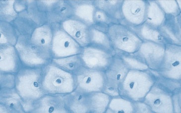
Bright field imaging provides a wealth of information for automated confluence determination, cell migration analysis and clone verification. (Spark Multimode Reader)
1. No more guessing
Whether out of convenience or necessity, it’s all too common to rely purely on visual inspection with a manual microscope to check on cell health and estimate confluence of adherent monolayers prior to running a microplate assay. Not only does this ‘guesstimation’ make for unreliable quality control, it is also time-consuming and disruptive to move between the microscope and your plate reader. On-board cell counting and confluence analysis provide rapid results, eliminate subjectivity, and give you images as documentary evidence that you can store for future reference. And you needn’t give up manual control altogether – a good imaging microplate reader keeps you in the driver’s seat with full control to focus, view and navigate around the well or slide, just as you would on a manual microscope.
2. Improve assay consistency
Cell confluence levels can significantly affect signaling pathways, proliferation rates and responsiveness to various treatments. For this reason, variability in cell confluence at the start of your assay can translate into misleading results and unacceptably large CVs. You can avoid these pitfalls by starting your experiments at the optimal percentage confluence every time. To achieve this, look for an imaging plate reader with responsive control software that can accurately measure cell confluence and then automatically initiate reagent addition or imaging at the confluence threshold of your choice.
3. Ditch your BCA assay
Normalization of assay signal to percentage confluence is becoming the “New Normal” for high quality plate reader assays. Forget destructive protein assays, additional dyes and recombinant fluorescent proteins as surrogates for cell mass determination. With bright field confluence measurements, you don’t waste valuable time, risk introducing artifacts or otherwise constrain your assay with cumbersome lysis protocols, unnecessary dyes or transfections. Image-based confluence assessment is a label-free, non-destructive approach that can be as accurate for assay normalization as conventional protein stains or fluorescent proteins (Figure 1). What’s more, imaging can give you important information about cell distribution that you can’t get from a protein assay. For example, it can clue you in to uneven seeding or edge-well effects that could be skewing your results.
A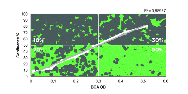

B 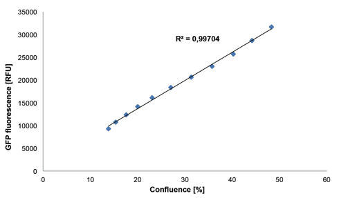

Figure 1: Image-based cell confluence determination is as accurate for assay normalization as protein staining (A) or fluorescent proteins (B), and provides additional spatial information. (Via Spark Multimode Reader).
4. Monitor proliferation - for free!
Automated confluence determination can also be a simple yet effective means of monitoring cell proliferation. This is information you get “for free” in addition to whatever else you are measuring in the same sample, because it doesn’t require any added fluorophores or markers. By avoiding the addition of potentially toxic dyes or other reagents used for conventional proliferation assays, you can monitor proliferation in real-time over extended periods. With a multimode reader, you can complement label-free proliferation assays with additional end-point or live-cell indicators (e.g., for ATP, caspase activity, etc.) for even more in-depth characterization. Since proliferation data will ideally be gathered over several days, reliable on-board environmental control is also a key consideration for success.
5. Do More, Discover More
Imaging capability integrated into a multimode plate reader gives you added power to drive your research in new directions, exploring the exciting world of live cell imaging while still running all the core assays you’ve come to rely on. Live-cell imaging opens the door to kinetic analysis of dynamic processes such as cell migration and wound healing. With whole-well bottom-read imaging capability, you can automate cell colony identification and verify clonality, which can be essential for applications such as therapeutic antibody development. Even more exciting is the potential to combine cell imaging with diverse absorbance, fluorescence and luminescence assays, opening up a wealth of completely new possibilities for your research.
Download our application notes learn more about the power of on-board bright field imaging for automated confluence determination, cell migration analysis and clone verification.

About the author

Christian Oberdanner
Dr. Christian Oberdanner is Product Manager at Tecan Austria. He studied molecular biology at the University of Salzburg with a strong focus on cell- and tumor biology during his PhD study. Christian started to work for Tecan Austria as external scientific consultant in 2005 and permanently joined the company in 2006. Since then he held the role of an Application Specialist in the research and development department as well as in the sales and marketing department and since 2015 of a Product Manager. Christian’s priority within Tecan is the multimode microplate reader portfolio.