


By Dr Christian Oberdanner
As we saw in the previous article in this series, detecting differences in your cell-based fluorescence experiments means you need high assay sensitivity and reproducibility that comes from high quality optics and intelligent measurement methods. All this can be achieved using Spark™ multimode microplate reader.
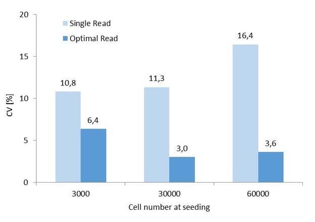
Optimal Read gives much higher reproducibility (low Coefficient of Variation) compared to Single Read (48-well plate).
Fusion Optics quickly get you onto the right wavelength
Selecting the optimal wavelengths for excitation and emission is fundamental to success in fluorescence assays. Multimode readers can include filters or monochromators (MCRs) to select excitation and emission wavelengths in fluorescence applications. MCRs enable you to select the exact wavelength desired and will give you excellent flexibility for a variety of applications, and only MCRs enable precise spectral scans in small steps. They do, however, have a lower light transmission than filters, which reduces sensitivity. Filter-based readers do offer higher sensitivity, but they also limit you to pre-defined wavelengths and are therefore only suitable for certain assays.
Hybrid systems offer the flexibility of both MCRs and filters, but have been limited to the selection of MCRs OR filters in one experiment. So assay development has still been a compromise. Until now. The unique Fusion Optics of Spark multimode microplate reader enable you to combine MCRs with filters in the same experiment to get truly optimal wavelength selections for maximum sensitivity. This means that it is now possible to measure with filter on excitation and MCR on emission site or vice versa. This unique ability to combine the flexibility of MCRs with the sensitivity of filters is especially relevant for fluorescence emission or excitation scans. And the new SparkControl™ software makes selecting the MCR or the wavelength filters easy.
Get focused. And stay that way.
Armed with optimal excitation and emission wavelengths, the next step is to make sure that you are measuring signal from the right place. The automated Z-focusing of the Spark reader automatically determines the focal point for the best signal-to-blank ratio in each microplate well, regardless of plate type, well shape or sample volume. This means that you can be sure that you are getting the best sensitivity by measuring signal from just your cell layer, and avoiding background from the medium. In every well. On every plate.
Maximize coverage. Efficiently.
You are now on track to getting high sensitivity fluorescence measurements specifically from the cell layer in the culture wells. But you also need to ensure that you are measuring over the whole culture surface since many cell types tend to grow unevenly over the bottom. Many plate readers offer the possibility of running multiple reads per well, but this generally demands user input and patterns will differ between plate types. Added to that, the flash number is often fixed, which means that increasing the coverage of the well bottom can be very time consuming.
The unique Optimal Read Function of Spark, however, ensures you get rapid and excellent coverage of cell cultures. The well is illuminated uniformly during excitation and whole-well emission. This ensures that even heterogeneous cell layers can be analyzed reliably and reproducibly by making multiple measurements on spots arrayed over each well. The process is rapid since the flash number is automatically distributed between the measurement spots. The result is a single value for every well, from 6- to 384-well plates, with a reproducibility that can give up to a four-fold improvement in the coefficient of variation.
Normalize on confluence level
Now is the time to add the final polish to your data by normalizing using cell confluence levels to spot outliers and anything else that might confound the interpretation of your results. The cell-imaging module of Spark enables you to precisely determine the confluence level in a well from 10 to 90 %, which greatly increases the quality of your data. And combined with the Optimal Read Function, this confluence analysis means you get close to an imaging system.
A flexible multimode microplate reader that boosts assay performance
Spark multimode microplate reader also has a number of other functions. High definition well scans give you a qualitative image of the fluorescently labeled cell population in a microplate, or you can create a low-resolution fluorescence image of you microplate well. Environmental control ensures that your cell cultures remain healthy even during lengthy experiments, and you can monitor and analyze your cells by viability analysis, and live cell imaging.
Whatever you need to do, Spark multimode plate reader will bring your fluorescence cell-based assays to a new level.

Keywords:
About the author

Christian Oberdanner
Dr. Christian Oberdanner is Product Manager at Tecan Austria. He studied molecular biology at the University of Salzburg with a strong focus on cell- and tumor biology during his PhD study. Christian started to work for Tecan Austria as external scientific consultant in 2005 and permanently joined the company in 2006. Since then he held the role of an Application Specialist in the research and development department as well as in the sales and marketing department and since 2015 of a Product Manager. Christian’s priority within Tecan is the multimode microplate reader portfolio.