


Organoids are three-dimensional (3D) cell culture systems that replicate key structural and functional aspects of human tissues. Derived from stem cells, organoids can self- organize and differentiate in vitro, mimicking the architecture and physiology of native organs. Their development has significantly advanced the field of cell biology by enabling researchers to study complex biological processes in controlled, human- relevant systems. The physiological relevance of organoids allows for more predictive and ethically sustainable research approaches, especially in the fields of drug development and personalized medicine.
Founded in 2013 in Leiden in the Netherlands, MIMETAS is an innovative Biotechnology company that focuses on development of 3D human relevant tissue models for drug discovery and drug development. They strive to develop robust, screenable organ models utilizing their versatile technology the OrganoPlate® platform. Partnering with many of the world’s leading pharmaceutical companies, MIMETAS now has over 80 employees, spread over four sites in the Netherlands, the USA and Japan.
One of the key challenges in organoid culture that MIMETAS is working to address is the improvement of reproducibility, particularly during the pre-culture phase when stem cell-derived cells are beginning to self-organize into complex 3D structures. At this stage, researchers usually have to rely on visual inspection by eye to assess growth, morphology, and formation success. However, this approach is subjective, prone to variability and lacks reproducibility, especially during scale-up. These limitations have driven the need for more quantitative, automated methods to assess organoid formation and quality in situ.
To enhance reproducibility in organoid-based models, MIMETAS has established standardized operating procedures (SOPs) that define key handling steps and culture timelines. These protocols are supported by routine quality-control measures, including the evaluation of critical reagents such as the gel matrix, media components and organoid batches.
As part of this workflow, the MIMETAS team is using the Tecan Spark® Cyto to enable both visual comparison and quantitative monitoring of organoid cultures. Parameters such as fragment size, morphology and growth behavior are assessed to support consistent outcomes. Notably, fragment size immediately after splitting was found to influence early growth dynamics in a donor-dependent manner. By leveraging the Spark’s imaging capabilities, the fragment sizes are quantified across samples and donors, generating insights into how initial size distributions affect subsequent organoid development. This data-driven approach also facilitates batch-to-batch comparison and helps identify potential inconsistencies arising from reagent variability or deviations in handling.

Longitudinal monitoring of organoid cultures showed that different production rounds exhibited consistent growth patterns, indicating improved control over culture conditions, while measuring organoid size immediately after splitting also allowed for estimation of population doubling times. In one colon organoid donor, larger fragments were linked to slower proliferation, while in other donors, initial fragment size had minimal effect on doubling time but appeared to limit overall growth and final organoid size. These findings underscore the donor-specific influence of fragment size on growth behavior.
Beyond physical parameters, media composition – particularly the use of growth versus differentiation conditions – also impacted organoid performance. Standardizing initial conditions, such as fragment size and seeding density, was found to improve consistency during both expansion and differentiation phases. Real-time monitoring with the Tecan Spark® Cyto supported this optimization by providing continuous insight into organoid responses under varying media conditions, facilitating more reproducible and controlled culture workflows.
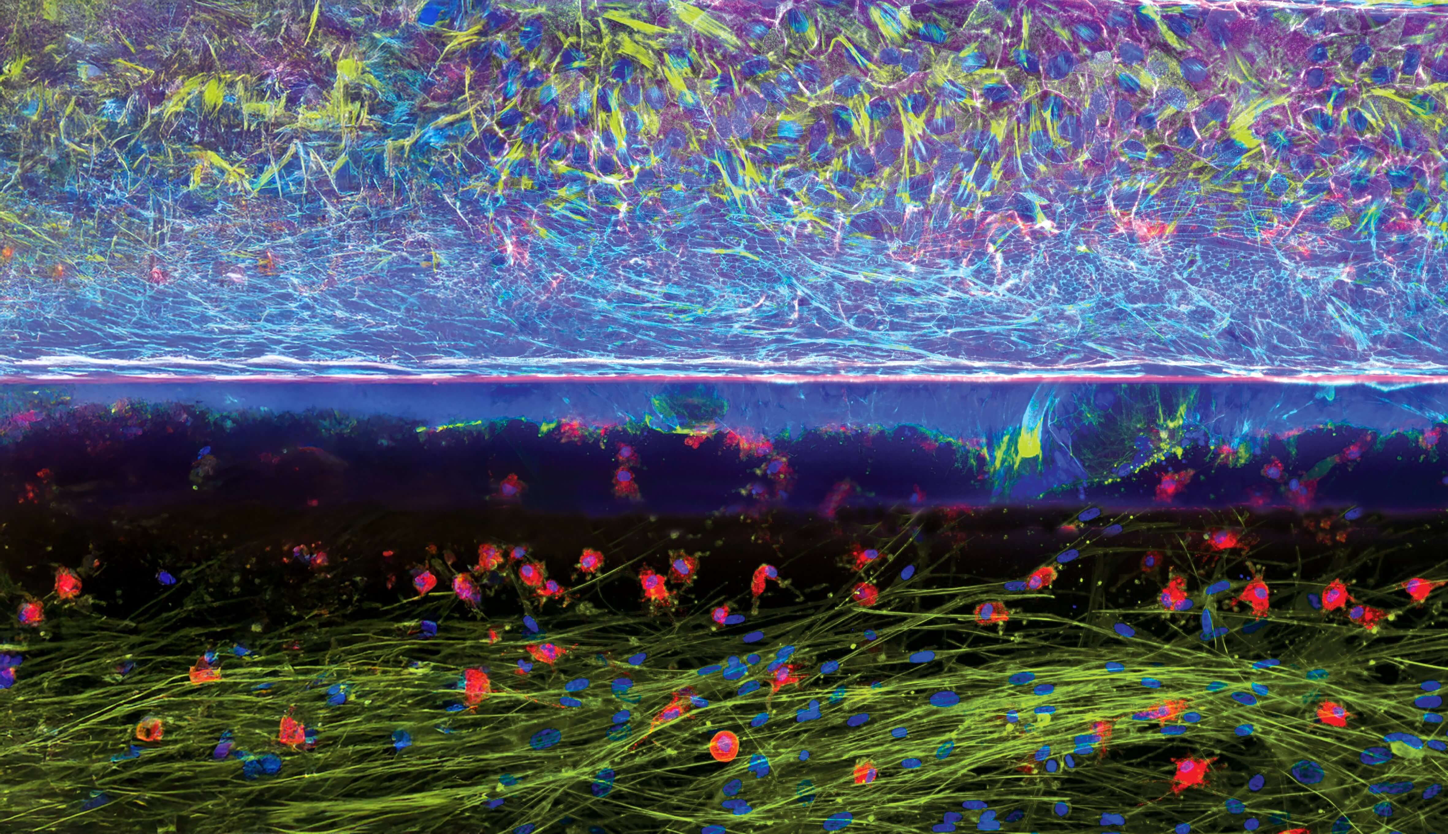
 Figure 1. Monitoring organoid cultures using the Tecan Spark® Cyto’s 3D imaging capabilities.
A) Representative image of an organoid droplet acquired three days after splitting. Yellow outlines indicate organoids detected automatically by the imaging software.
B) Quantification of organoid number on day 3 post-seeding across multiple passages and donor-derived organoid banks, demonstrating consistent detection and culture behavior.
C) Measurement of mean organoid area immediately after splitting, illustrating how initial fragment size influences subsequent growth dynamics and population doubling behavior (D).
MIMETAS has used these optimized pre-culture conditions to develop assay-ready organoids for reproducible research, including the OrganoReady® Colon Organoid model. Derived from human adult stem cells, the organoids are expanded under controlled conditions and seeded into the OrganoPlate® platform, a microfluidic system that supports membrane-free, perfused culture of 64 organoid tubules under physiological conditions. Delivered to researchers’ labs, fully optimized and ready-to- use, the OrganoReady Colon Organoid enables scientists to focus directly on assay execution, supporting applications in drug screening and toxicology and offering a consistent and reliable colon organoid model for reproducible research outcomes.
The partnership with MIMETAS is a good example of how Tecan contributes, together with partners, to scaling healthcare innovation in this exciting field. Multimodal plate readers such as the Tecan Spark® Cyto equipped with 3D live-cell imaging modules enable non-invasive, real-time monitoring of key culture parameters such as growth dynamics, seeding density and fragment size. When combined with standardized protocols and microfluidic platforms like the OrganoPlate® from MIMETAS, this integrated approach supports more consistent organoid expansion and differentiation.
As these technologies continue to advance, they will play a central role in scaling organoid workflows with greater control and reliability, contributing towards the development of new, more effective treatments for challenging diseases and ultimately improving outcomes for patients worldwide.
Spark Cyto is for research use only. Not for use in diagnostic procedures.