


Many of the anti-cancer drugs currently used for chemotherapy work by causing replication-associated DNA damage that kills individual cancer cells. While this can be an effective way of treating the disease, the drugs often also indiscriminately affect healthy cells, causing unpleasant side effects for the patient. To help resolve this problem, researchers at the Karolinska Institute in Sweden are developing DNA repair inhibitors that are able to selectively introduce toxic DNA damage to cancer cells, while avoiding causing harm to normal cells, to support the successful treatment of cancers and improving patients’ experience of chemotherapy.
The Helleday Lab sits within the Department of Oncology-Pathology at the Karolinska Institute, and primarily investigates basic DNA repair, DNA damage signaling pathways and nucleotide metabolism. The lab’s overarching aim is to develop novel targeted therapies for cancer, inflammation and autoimmunity, as well as viral infections. The multidisciplinary team was among the first to demonstrate the synthetic lethality concept for the treatment of cancer, exploiting the high level of DNA damage and dysfunctional redox status present in cancer cells to prevent the repair of lesions and stimulate further injury to the cell. The advantage of this approach is that these treatments should have far fewer negative side effects on patients than existing chemotherapies.
 The Thomas Helleday Research Team
Dr Oliver Mortusewicz is the Principal Researcher and Basic Science Team Leader of the Thomas Helleday Research Team within the lab. He described the crucial work carried out by his group: “Our goal is to turn cancer defects into cures by targeting altered DNA repair and cellular metabolism found in cancerous cells. We aim to take our projects all the way from basic science to the clinic, and we have already been successful, with two Phase I clinical trials currently being conducted using our MTH1 inhibitor candidate – karonudib/OXC-101 – at Karolinska University Hospital.”
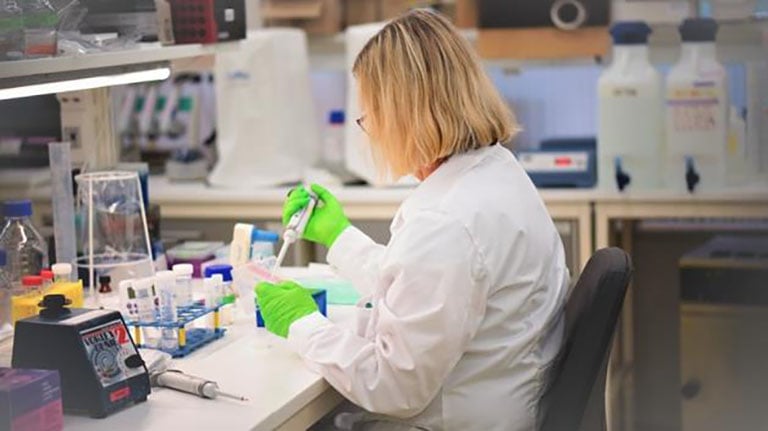
The lab performs a wide range of tests as part of its drug discovery and development workflow, including target engagement assays to ensure the candidate molecule is binding to the target in the cancer cell, functional assays to test for the efficacy of drugs that induce DNA damage, and synergy assays to investigate how its potential anti-cancer compounds work synergistically with approved drugs. For all of these assays, the team needs to accurately dispense microliter, nanoliter, or even picoliter volumes of both candidate and approved therapeutics into 384-well plates prior to seeding the plates with cancer cells.
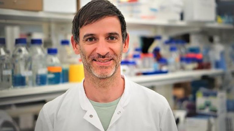 Dr Oliver Mortusewicz
This low volume dispensing is difficult to do by hand accurately at the scale required, and so the lab relies on a D300e Digital Dispenser to provide non-contact dispensing down to 11 pl. Before acquiring the D300e platform, staff had to perform these complex and extensive pipetting tasks manually, making them extremely time consuming, tedious and prone to human error. Oliver explained the difference the instrument has made to the lab’s productivity: “The D300e makes our lives so much easier, improving our efficiency and throughput by speeding up our liquid handling workflow and reducing hands-on time. In turn, this frees up the staff to do other vital tasks in the lab. Another significant benefit of the D300 is that it reduces the possibility of errors by automating many of the pipetting steps, which enhances our assay reproducibility.”
Our goal is to turn cancer defects into cures by targeting altered DNA repair and cellular metabolism found in cancerous cells.
Oliver continued: “We also conduct resazurin assays to test for cell viability, but this test depends on the metabolic activity of the cells, which doesn’t correlate 100 % with cell viability. That’s why we also subsequently image the plates with a Spark® Cyto imaging plate reader. This image-based analysis allows us to get a better understanding of what is actually happening to the cells through the use of fluorescently tagged proteins or immunofluorescence. For example, we can gauge the rate of proliferation or the localization of repair proteins upon the induction of DNA damage. A major advantage of the Spark Cyto over a classical plate reader is its ability to perform real-time imaging and cytometry. For example, we can carry out automated stain-free cell counting and confluence assessment of a whole microplate in just one image per well using the system’s bright field imaging technology.”
 Researchers are using DNA repair inhibitors to selectively introduce toxic DNA damage to cancer cells
Oliver and his colleagues now rely on the two Tecan platforms for many of their workflows, and are looking forward to employing them in further vital research going forward: “Both of the platforms are so easy to learn, and the interface is very user friendly. We have many students working in the lab with us and it doesn’t take long to train them up on it. That means they can get started on experiments sooner, which is great for our assay productivity. In addition, we have discovered that one of our anti-cancer molecules is actually also active in preventing inflammation, so we are now venturing out into other areas of research with our drugs, following wherever the science leads us. We certainly plan to keep using our versatile Tecan platforms in the future to support us in our upcoming investigations,” Oliver concluded.
For research use only. Not for use in clinical diagnostics.