


By Oliver Schmidt
sIL-2 receptor: a biomarker for immune activation
The quantification of soluble interleukin-2 receptor (sIL-2R) in serum or plasma in adults has become an extremely useful tool for clinicians to assess immune function in vivo, as part of the investigation, management, or prognosis of a broad spectrum of diseases. However, the measurement of sIL-2R is simple compared to the complex story behind this biomarker that has become so central to our study of immune-mediated disease.
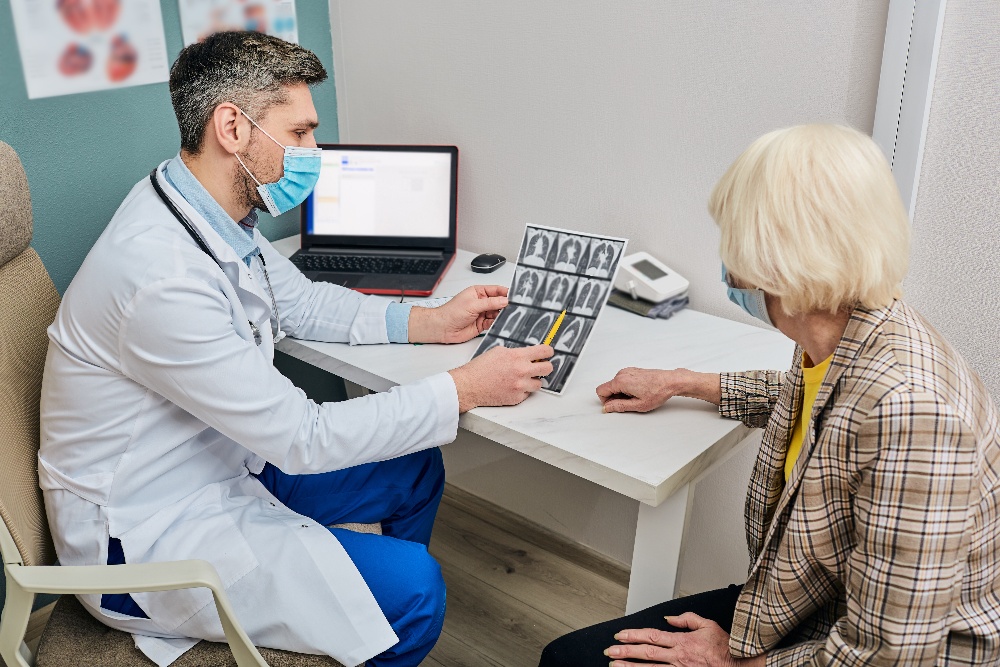
Doctor analyzing a CT scan of a patient’s lungs. Around 90% of sarcoidosis patients have lung symptoms, with elevated sIL-2R levels indicating immune activation.
Why measure soluble interleukin-2 receptor levels?
Interleukin-2 (IL-2) is a key signaling molecule in the human immune system. It is a cytokine, one of a group of small, secreted proteins released by cells that have a specific effect on the interactions and communication between cells. As such, IL-2 regulates the activities of the white blood cells that are responsible for immunity, forming part of the body's natural response to infection, and helping it to discriminate between foreign ("non-self") and "self".1
IL-2 mediates its effects by binding to IL-2 receptors, which are expressed by lymphocytes. The major sources of IL-2 are activated CD4+ T cells and activated CD8+ T cells.1 On T-cell activation, the soluble form of the interleukin-2 receptor (sIL-2R) is secreted, or “shed”, meaning that elevated concentrations of sIL-2R are found in patients suffering from any one of an extensive range of conditions that are associated with an ongoing immune response.1
The immune-mediated diseases associated with increased levels of sIL-2R include but are not limited to sarcoidosis, multiple sclerosis, biliary cirrhosis, chronic immune activation in common variable immunodeficiency (CVID) and hemophagocytic lymphohistiocytosis (HLH).2-6
The convenient implication of this association is that sIL-2R could hypothetically be used as a generic biomarker to monitor/predict disease activity and treatment response that is specific to these diseases, and indeed many more.7,8 However, to be certain we are measuring something that is clinically relevant, requires us to delve a little deeper into how the IL-2 / sIL-2R system functions.
To deepen your understanding of sIL-2R and its use in the laboratory, watch a webinar featuring Dr. Bob Meek from St. Antonius Hospital, providing valuable insights into the subject.

IL-2 and sIL-2R in immune activation: unraveling the twisted plot
Binding of IL-2 to sIL-2R can either reduce or enhance immune response, depending on the target cell being involved in immunity or self-tolerance.9,10 The history of the so-called soluble interleukin-2 receptor — variously termed sIL-2R, sCD25, sTAC, sIL2R, IL-2RA — goes back to 1985, when it was first described as being actively released by activated peripheral blood T-cells via proteolytic cleavage of the cell surface IL-2R.11
In 1990, Rubin and colleagues were the first to demonstrate that after in vitro activation, T lymphocytes not only enhanced cellular IL-2R expression but also released soluble IL-2R(α), and that, similarly to cellular IL-2R expression, the release of soluble IL-2R required de novo protein synthesis rather than cellular proliferation.12
However, despite this long-recognized association between immune activation and increased sIL-2R release under pathological conditions, the biological actions of sIL2R are still far from understood. Several mechanisms of action, ranging from immune-inhibitory to immuno-stimulatory effects, have been proposed. Figure 1 illustrates a little of the paradox and complexity of the IL-2 / sIL-2R system.10
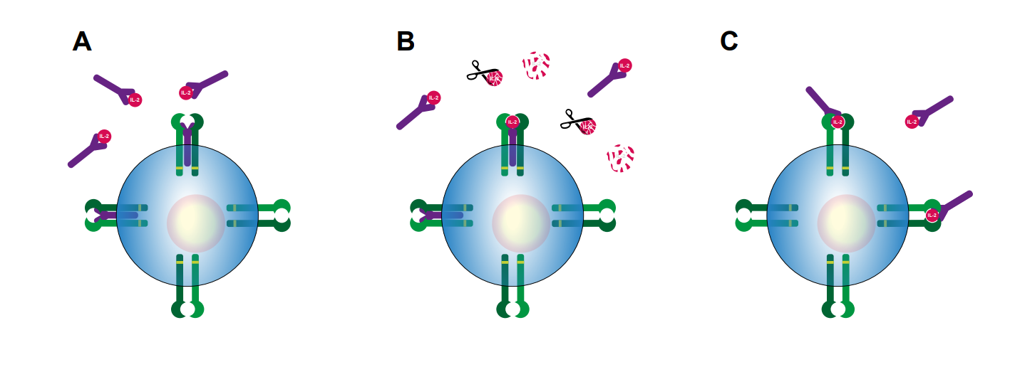
Figure 1. Potential mechanisms of action of sIL-2R during immune activation, as proposed by Dik & Heron (2020).10 A) sIL-2R binds IL-2, thereby prohibiting IL-2 from binding to either to the low affinity dimeric IL-2 receptor or high affinity trimeric IL-2 receptor. B) sIL-2R binds IL-2, thereby protecting IL-2 from enzymatic degradation and prolonging IL-2 half-life. C) sIL-2R binds IL-2, thereby increasing the affinity of IL-2 for the low-affinity dimeric IL-2 receptor. IL-2Rα: purple, IL-2Rβ: light green, IL-2Rγ: dark green, sIL-2R: purple.
Soluble IL-2R binds IL-2 efficiently, and based on in vitro experiments, it has been proposed that sIL-2R may limit activation and proliferation of T lymphocytes by sequestration of available IL-2.13 However, conflicting data have been reported.14 Alternatively, sIL-2R complexed with IL-2 may prolong IL-2 half-life, possibly enhancing the immune-stimulatory properties of IL-2, even by activation of low-affinity dimeric IL-2R.15
It is possible that IL-2 can be presented to CD4+ T lymphocytes through sIL-2R, which then induces differentiation into Tregs (rather than differentiation into T-helper (Th)1 or Th17 lymphocytes) that subsequently can suppress immune activity.16 On the other hand, there are reports to support observations that sIL-2R may promote (auto)immune processes in association with enhanced Th17 generation, which involves sequestration of the IL-2 that normally inhibits early Th17 differentiation.17
Despite the obvious complexity and lack of agreement so far regarding the exact mechanism(s) of action of different configurations of sIL-2R, as well as their in vivo occurrence and final biological effects, the data available so far do at least support a role for sIL-2R in regulating IL-2-dependent cell function.
What makes sIL-2R an appealing biomarker?
We saw earlier that increased blood levels of sIL-2R are considered to indicate an ongoing immune response, which could theoretically be used to monitor a huge range of immune-mediated diseases, which is a good starting point, even if the generic nature of the response might initially have been a concern.9
Generally speaking, conditions that are characterized by excessive production of lymphocytes, so-called lymphoproliferative disorders, show high sIL-2R levels compared to healthy controls, as do granulomatous diseases, such as sarcoidosis, in which T-cell activation is a typical hallmark.
The relative stability of sIL-2R levels throughout adult life, and the minimal gender-related differences are further useful attributes that help make sIL-2R an attractive biomarker.12,18 That said, individual differences in age, gender, lifestyle and general health may need to be taken into account when considering reference values and ranges.19 An indication of the levels of sIL-2R seen in healthy and diseased individuals can be seen in Table 1.
In the absence of a World Health Organization (WHO) standard, the measurements given in this table are assay-dependent, although it is recommended that kits are calibrated against the international reference standard NIBSC 97/600. However, when looking at the relative levels of sIL-2R, a fair conclusion is that in diseased individuals, sIL-2R levels are elevated compared to those in healthy populations (see Table 1).
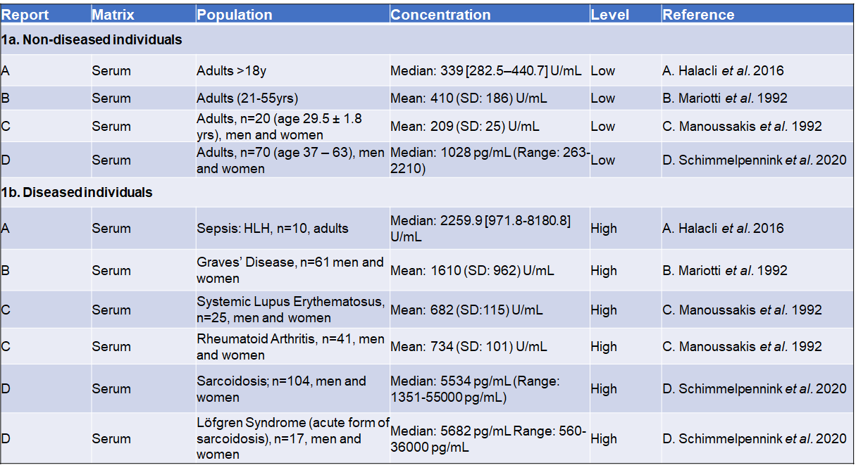
Table 1. Comparing levels of sIL-2 receptor in healthy and diseased individuals. A: Reference ranges for sIL-2R in an apparently healthy population measured in serum and plasma in adults. B: sIL-2R values for affected population measured in serum and plasma.
How do we measure soluble interleukin-2 receptor?
Soluble IL-2 receptor levels are generally measured using immunoassays, either enzyme-linked immunosorbent assay (ELISA), or chemiluminescent immuno-assay (CLIA). There is very good correlation between the results of both types of assays, although absolute values of ELISA (pg/mL) are about 7 – 8 times higher than those obtained by CLIA (U/mL). Commercially available ELISAs should be calibrated against the international reference standard NIBSC 97/600.
ELISA assays for sIL-2R are typically designed for batch analysis and have a measuring range up to 10,000 – 20,000 pg/mL, based on a standard serum dilution of ~1:5. Further dilutions will of course enable the quantification of higher sIL-2R levels. This can be facilitated by the use of automated liquid-handling systems, so that multiple dilutions can be analyzed simultaneously.
In clinical practice, laboratories will need to use different criteria for determining the upper limit of normal (cut-off) for sIL-2R levels: there is such a broad range of values (see Table 1) that distinct cut-off values may be required for each situation. These will depend on the disease that is being diagnosed or monitored, and on the stage at which sIL-2R levels are measured for that disease.
The principal characteristics of sIL-2 kits might include, but not be limited to:
- All kit reagents should be ready-to-use – no dilution necessary
- Include ready-to-use standard-curve reagents – no stock standard to dilute
- Kit ready-calibrated against NIBSC 97/600 Standard Preparation
- Internal kit controls provided
- Easily automatable
There are many Research Use Only (RUO) ELISA kits available for measuring sIL-2R levels. However, to be able to use a kit for diagnostic purposes it must comply with applicable regulations. In the European Union, this means that new launched kits used for IVD must conform to IVDR (Regulation (EU) 2017/746 for in vitro diagnostic medical devices). To this end, TECAN has recently developed an IVDR-compliant soluble Interleukin-2-Receptor ELISA Kit.
Explore the requirements, challenges, and available solutions for measuring soluble interleukin-2 receptor in a webinar with Dr. Bob Meek from St. Antonius Hospital.
Sarcoidosis and sIL-2R: a deep dive
Sarcoidosis, also known as Besnier-Boeck-Schaumann disease, is a rare condition that causes small patches of swollen tissue, called granulomas, to develop in the organs of the body.24 It often affects the lungs and lymph nodes and can also affect the skin. The symptoms of sarcoidosis depend on which organs are affected, but typically include tender bumps on the skin, shortness of breath, and a persistent cough.
It is impossible to predict how sarcoidosis will affect a person, as the condition can affect any organ and the symptoms vary widely depending on which organs are involved. Sarcoidosis may be acute or chronic, and some people do not have any symptoms at all, so that their condition might only be diagnosed after an X-ray carried out for another reason.
Elevated sIL-2R levels have been reported in numerous studies in sarcoidosis patients, and as such, sIL-2R is already an established biomarker for the disease.2,25 Some studies suggest the measurement of sIL-2R as a marker of therapy success.26 Sensitivity of serum sIL-2R as a diagnostic biomarker for sarcoidosis lies around 79%, and in patients with uveitis the sensitivity of elevated sIL-2R levels to establish underlying sarcoidosis is around 81%–98%, with an AUC of 0.76 (fair) and 0.96 (excellent). 27
Interestingly, patients with extrapulmonary involvement have been shown to have relatively high levels of serum sIL-2R, suggesting a value as staging and/or severity biomarker. Patients with more advanced radiographic stages and progressive disease show higher levels of sIL-2R levels. Serum sIL-2R tests have the highest ability to determine pulmonary severity in comparison to soluble angiotensin-converting enzyme (sACE). Furthermore, in contrast to sACE, an advantage to the measurement of sIL-2R levels is that the interpretation of serum sIL-2R levels is not confounded by the use of drugs or immunosuppressants.27
When using sIL-2R levels as a biomarker for sarcoidosis, it should be noted that an increase of serum sIL-2R values is not always disease-specific: elevated values can also be found in other conditions, including hematologic malignancies, other granulomatous diseases, various autoimmune disorders, and post transplantation. Renal insufficiency may also have a major impact on sIL-2 levels, which could lead to misinterpretation of test results.27
However, even given these caveats, and especially when considering the broader clinical picture of the individual patient, serum sIL-2R measurement is a very useful prognostic marker. High serum sIL-2R levels can predict the need for therapy in sarcoidosis patients, and high sIL-2R at initiation of therapy has even shown value as a predictor of relapse after therapy with infliximab.27
Changes in concentration of serum sIL-2R have been shown to be related to clinical changes, correlating well with changes in pulmonary function parameters and radiological abnormalities, and finally, the serial measurement of serum sIL-2R during disease follow-up has also proven useful to assess the evolution of disease activity in sarcoidosis.27 In conclusion, when it comes to establishing a credible tool for diagnosing, monitoring and treating sarcoidosis, sIL-2R measurement is here to stay.
The future of sIL-2R measurement
We have seen that soluble interleukin-2 receptor (sIL-2R) measurement can be an extremely useful tool for clinicians to assess immune function in vivo, across a broad spectrum of diseases, and established its key role in the investigation and management of sarcoidosis. Until recently, the use of sIL-2R measurement as standard in the clinical environment depended upon the development of IVDR-compliant assays. To this end, Tecan is now able to offer the first IVD-compliant sIL-2R ELISA for use in the diagnosis and therapeutic monitoring of autoimmune disease, paving the way for the broad adoption of sIL-2 ELISA in the clinic.
Disclaimers:
Product availability and regulatory status may vary across regions outside the EU depending on local country-specific regulation. Please consult your local Tecan office for more information.
Find out more about the sIL-2R ELISA kits available from Tecan here.

References
1. Liao, W., Lin, J. X., & Leonard, W. J. (2011). IL-2 family cytokines: new insights into the complex roles of IL-2 as a broad regulator of T helper cell differentiation. Current opinion in immunology, 23(5), 598–604.
PubMed ID: https://pubmed.ncbi.nlm.nih.gov/21889323/
DOI: https://doi.org/10.1016/j.coi.2011.08.003
2. Thi Hong Nguyen, C., Kambe, N., Kishimoto, I., Ueda-Hayakawa, I., & Okamoto, H. (2017). Serum soluble interleukin-2 receptor level is more sensitive than angiotensin-converting enzyme or lysozyme for diagnosis of sarcoidosis and may be a marker of multiple organ involvement. The Journal of dermatology, 44(7), 789–797.
PubMed ID: https://pubmed.ncbi.nlm.nih.gov/28295528/
DOI: https://doi.org/10.1111/1346-8138.13792
3. Peerlings, D., Mimpen, M., & Damoiseaux, J. (2021). The IL-2–IL-2 receptor pathway: Key to understanding multiple sclerosis. Journal of Translational Autoimmunity, 100123.
PubMed ID: https://pubmed.ncbi.nlm.nih.gov/35005590/
DOI: https://doi.org/10.1016/j.jtauto.2021.100123
4. Barak, V., Selmi, C., Schlesinger, M., Blank, M., Agmon-Levin, N., Kalickman, I., Gershwin, M. E., & Shoenfeld, Y. (2009). Serum inflammatory cytokines, complement components, and soluble interleukin 2 receptor in primary biliary cirrhosis. Journal of autoimmunity, 33(3-4), 178–182.
PubMed ID: https://pubmed.ncbi.nlm.nih.gov/19846277/
DOI: https://doi.org/10.1016/j.jaut.2009.09.010
5. Litzman, J., Nechvatalova, J., Xu, J., Ticha, O., Vlkova, M., & Hel, Z. (2012). Chronic immune activation in common variable immunodeficiency (CVID) is associated with elevated serum levels of soluble CD14 and CD25 but not endotoxaemia. Clinical and experimental immunology, 170(3), 321–332.
PubMed ID: https://pubmed.ncbi.nlm.nih.gov/23121673/
DOI: https://doi.org/10.1111/j.1365-2249.2012.04655.x
6. Lin, M., Park, S., Hayden, A., Giustini, D., Trinkaus, M., Pudek, M., Mattman, A., Schneider, M., & Chen, L. (2017). Clinical utility of soluble interleukin-2 receptor in hemophagocytic syndromes: a systematic scoping review. Annals of hematology, 96(8), 1241–1251.
PubMed ID: https://pubmed.ncbi.nlm.nih.gov/28497365/
DOI: https://doi.org/10.1007/s00277-017-2993-y
7. Durda, P., Sabourin, J., Lange, E. M., Nalls, M. A., Mychaleckyj, J. C., Jenny, N. S., Li, J., Walston, J., Harris, T. B., Psaty, B. M., Valdar, W., Liu, Y., Cushman, M., Reiner, A. P., Tracy, R. P., & Lange, L. A. (2015). Plasma Levels of Soluble Interleukin-2 Receptor α: Associations With Clinical Cardiovascular Events and Genome-Wide Association Scan. Arteriosclerosis, thrombosis, and vascular biology, 35(10), 2246–2253.
PubMed ID: https://pubmed.ncbi.nlm.nih.gov/26293465/
DOI: https://doi.org/10.1161/ATVBAHA.115.305289
8. Karim, A. F., Eurelings, L., Bansie, R. D., van Hagen, P. M., van Laar, J., & Dik, W. A. (2018). Soluble Interleukin-2 Receptor: A Potential Marker for Monitoring Disease Activity in IgG4-Related Disease. Mediators of inflammation, 2018, 6103064.
PubMed ID: https://www.ncbi.nlm.nih.gov/pmc/articles/PMC5854105/
DOI: https://doi.org/10.1155/2018/6103064
9. Damoiseaux J. (2020). The IL-2 - IL-2 receptor pathway in health and disease: The role of the soluble IL-2 receptor. Clinical immunology (Orlando, Fla.), 218, 108515.
PubMed ID: https://pubmed.ncbi.nlm.nih.gov/32619646/
DOI: https://doi.org/10.1016/j.clim.2020.108515
10. Dik, W. A., & Heron, M. (2020). Clinical significance of soluble interleukin-2 receptor measurement in immune-mediated diseases. The Netherlands journal of medicine, 78(5), 220–231.
PubMed ID: https://pubmed.ncbi.nlm.nih.gov/33093245/
11. Rubin, L. A., Kurman, C. C., Fritz, M. E., Biddison, W. E., Boutin, B., Yarchoan, R., & Nelson, D. L. (1985). Soluble interleukin 2 receptors are released from activated human lymphoid cells in vitro. Journal of immunology (Baltimore, Md. : 1950), 135(5), 3172–3177.
PubMed ID: https://pubmed.ncbi.nlm.nih.gov/3930598/
12. Rubin, L. A., & Nelson, D. L. (1990). The soluble interleukin-2 receptor: biology, function, and clinical application. Annals of internal medicine, 113(8), 619–627.
PubMed ID: https://pubmed.ncbi.nlm.nih.gov/2205142/
DOI: https://doi.org/10.7326/0003-4819-113-8-619
13. Rubin, L. A., Jay, G., & Nelson, D. L. (1986). The released interleukin 2 receptor binds interleukin 2 efficiently. Journal of immunology (Baltimore, Md. : 1950), 137(12), 3841–3844.
PubMed: https://pubmed.ncbi.nlm.nih.gov/3023487/
14. Pedersen, A. E., & Lauritsen, J. P. (2009). CD25 shedding by human natural occurring CD4+CD25+ regulatory T cells does not inhibit the action of IL-2. Scandinavian journal of immunology, 70(1), 40–43.
PubMed: https://pubmed.ncbi.nlm.nih.gov/19522766/
DOI: https://doi.org/10.1111/j.1365-3083.2009.02268.x
15. Vanmaris, R. M. M., & Rijkers, G. T. (2017). Biological role of the soluble interleukin-2 receptor in sarcoidosis. Sarcoidosis, vasculitis, and diffuse lung diseases : official journal of WASOG, 34(2), 122–129.
PubMed ID: https://www.ncbi.nlm.nih.gov/pmc/articles/PMC7170137/
DOI: https://doi.org/10.36141/svdld.v34i2.5369
16. Yang, Z. Z., Grote, D. M., Ziesmer, S. C., Manske, M. K., Witzig, T. E., Novak, A. J., & Ansell, S. M. (2011). Soluble IL-2Rα facilitates IL-2-mediated immune responses and predicts reduced survival in follicular B-cell non-Hodgkin lymphoma. Blood, 118(10), 2809–2820.
PubMed ID: https://www.ncbi.nlm.nih.gov/pmc/articles/PMC3172797/
DOI: https://doi.org/10.1182/blood-2011-03-340885
17. Maier, L. M., Anderson, D. E., Severson, C. A., Baecher-Allan, C., Healy, B., Liu, D. V., Wittrup, K. D., De Jager, P. L., & Hafler, D. A. (2009). Soluble IL-2RA levels in multiple sclerosis subjects and the effect of soluble IL-2RA on immune responses. Journal of immunology (Baltimore, Md. : 1950), 182(3), 1541–1547.
PubMed ID: https://www.ncbi.nlm.nih.gov/pmc/articles/PMC3992946/
DOI: https://doi.org/10.4049/jimmunol.182.3.1541
18. Taniguchi, T., & Minami, Y. (1993). The IL-2/IL-2 receptor system: a current overview. Cell, 73(1), 5–8.
PubMed ID: https://pubmed.ncbi.nlm.nih.gov/8462103/
DOI: https://doi.org/10.1016/0092-8674(93)90152-g
19. Vanessa Alende-Castro, Manuela Alonso-Sampedro, Carmen FernándezMerino, Bernardo Sopeña, Carmen Vidal, Francisco Gude & Arturo GonzalezQuintela (2023). Factors influencing serum concentrations of soluble interleukin-2 receptor: a general adult population study, All Life, 16:1, 2169958
DOI: https://doi.org/10.1080/26895293.2023.2169958
20. Halacli, B., Unver, N., Halacli, S. O., Canpinar, H., Ersoy, E. O., Ocal, S., Guc, D., Buyukasik, Y., & Topeli, A. (2016). Investigation of hemophagocytic lymphohistiocytosis in severe sepsis patients. Journal of critical care, 35, 185–190.
PubMed ID: https://pubmed.ncbi.nlm.nih.gov/27481757/
DOI: https://doi.org/10.1016/j.jcrc.2016.04.034
21. Mariotti, S., Caturegli, P., Barbesino, G., Marinò, M., Del Prete, G. F., Chiovato, L., Tonacchera, M., De Carli, M., & Pinchera, A. (1992). Thyroid function and thyroid autoimmunity independently modulate serum concentration of soluble interleukin 2 (IL-2) receptor (sIL-2R) in thyroid diseases. Clinical endocrinology, 37(5), 415–422.
PubMed ID : https://pubmed.ncbi.nlm.nih.gov/1486691/
DOI: https://doi.org/10.1111/j.1365-2265.1992.tb02352.x
22. Manoussakis, M. N., Germanidis, G. S., Drosos, A. A., & Moutsopoulos, H. M. (1992). Impaired urinary excretion of soluble IL-2 receptors in patients with systemic lupus erythematosus and rheumatoid arthritis. Lupus, 1(2), 105–109.
PubMed ID: https://pubmed.ncbi.nlm.nih.gov/1301961/
DOI: https://doi.org/10.1177/096120339200100208
23. Schimmelpennink, M. C., Quanjel, M., Vorselaars, A., Wiertz, I., Veltkamp, M., Van Moorsel, C., & Grutters, J. C. (2020). Value of serum soluble interleukin-2 receptor as a diagnostic and predictive biomarker in sarcoidosis. Expert review of respiratory medicine, 14(7), 749–756.
PubMed ID: https://pubmed.ncbi.nlm.nih.gov/32248706/
DOI: https://doi.org/10.1080/17476348.2020.1751614
24. https://www.nhs.uk/conditions/sarcoidosis/ Accessed 13 April 2023 25. Eurelings, L. E. M., Miedema, J. R., Dalm, V. A. S. H., van Daele, P. L. A., van Hagen, P. M., van Laar, J. A. M., & Dik, W. A. (2019). Sensitivity and specificity of serum soluble interleukin-2 receptor for diagnosing sarcoidosis in a population of patients suspected of sarcoidosis. PloS one, 14(10), e0223897.
PubMed ID: https://pubmed.ncbi.nlm.nih.gov/31622413/
DOI: https://doi.org/10.1371/journal.pone.0223897
26. Vorselaars, A. D., van Moorsel, C. H., Zanen, P., Ruven, H. J., Claessen, A. M., van Velzen-Blad, H., & Grutters, J. C. (2015). ACE and sIL-2R correlate with lung function improvement in sarcoidosis during methotrexate therapy. Respiratory medicine, 109(2), 279–285.
PubMed ID: https://pubmed.ncbi.nlm.nih.gov/25496652/
DOI: https://doi.org/10.1016/j.rmed.2014.11.009
27. Milou C. Schimmelpennink MD, Adriane D.M. Vorselaars MD, PhD, Jan C. Grutters MD. Sarcoidosis: A Clinician's Guide (2019), Chapter 19 - Biomarkers in Sarcoidosis. Pages 219-238.
PubMed ID: https://pubmed.ncbi.nlm.nih.gov/25496652/
DOI: https://doi.org/10.1016/B978-0-323-54429-0.00019-7
Keywords:
About the author

Oliver Schmidt
Oliver Schmidt is product manager for research reagents, neurodegeneration, and bone and mineral products at TECAN-IBL in Hamburg with more than 10 years’ experience in bringing these products to the Life Science and academic community. He studied Environmental Engineering at the Technical University of Hamburg-Harburg and joined TECAN-IBL in 2005.