


Damage to the brain or spinal cord can be life changing for affected individuals, and it was historically thought that these injuries would not heal and could not be repaired. However, since the discovery of neurite growth inhibitors by Professor Martin E. Schwab at the University of Zurich, clinical researchers have been exploring new therapeutic approaches to treat cerebral stroke and spinal cord injury. The Wyss Zurich/University of Zurich CeNeReg project and NovaGo Therapeutics Inc. – co-founded by Professor Schwab – are at the forefront of this exciting field, and are dedicated to the development of human antibody therapeutics to stimulate nerve repair and regeneration.
The concept of neurite growth inhibitors as the key reason for the lack of regeneration in the central nervous system was first proposed by Professor Schwab in the early 1990s. His team at the University of Zurich then went on to discover a key inhibitory factor – Nogo-A – which it identified as a potential therapeutic target that could be inhibited to allow neurons damaged by cerebral stroke or spinal cord injury to regrow and form new networks. Since this time, a large number of preclinical studies have been performed by independent laboratories around the world, validating the unique potential of anti-Nogo-A antibodies as a new therapeutic approach. Start-up biotech NovaGo Therapeutics and Wyss Zurich – a foundation supporting translational medicine within the University of Zurich and ETH Zurich – are determined to translate this knowledge from a research to a clinical environment, with the aim of developing novel immunotherapies that would enable the regeneration of disrupted nerve tissue.

Michael Maurer (back row, second from right) and the NovaGo team relaxing away from the lab
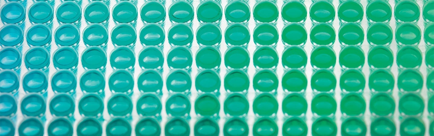
Dr Michael Maurer, a former senior scientist at Wyss Zurich and currently a senior scientist at NovaGo, explained: “We are developing and characterizing an anti-Nogo-A antibody for health authorities, and needed a system that would allow in-depth characterization of these complex macromolecules. Many of our assays are cell- or ELISA-based, making a high performance plate reader essential. I initially began looking for a system to perform ELISAs, but quickly realized that a multimode system that could also quantify DNA, perform fluorescence measurements, and count and image cells, would be the best solution. The Spark seemed ideal, as it could do everything, so I asked Tecan for a demonstration, and was really surprised at just how good this system is.”
“Before getting the Spark, we had a number of benchtop instruments in the lab, but it has now replaced all of them. The space-saving is not so important for us, but only having a single instrument to maintain is an important saving. The only downside of having an instrument that does everything well is that a lot of people want to use it! Fluorescence and absorbance measurements are very easy, and the imaging capabilities are good. One of the first things we used the system for was to take pictures of our cultures during cell-based assays, and this worked really well straightaway. We also use this function to perform cell adhesion assays, as Nogo-A – either fragments or total protein from tissue samples – blocks cell adhesion.”
I t’s very easy to develop new methods; everything is very intuitive and self-explanatory – I’ve never even opened the manual!
Most multimode readers can be equipped with either filters or monochromators (MCRs) to define excitation and emission wavelengths in fluorescence applications, but Spark is equipped with both, allowing the user to independently choose between filters and MCRs for both excitation and emission. Michael illustrated the value of this: “All the assays we run on the Spark are developed and validated in house, and the flexibility of the MCRs is ideal for the development of fluorescence-based methods, speeding up the whole process. To have them for both excitation and emission is really awesome, because you are not limited by the filters that you have available. Then, once you’ve optimized your assay, you can switch to the more sensitive filters and create a standard curve to check the limits of detection and maximum quantification.”
Keywords: