


Chromosomal tagging using advanced fluorescent labeling techniques can help us to understand cells at a systemic level. Researchers in Germany have developed a universal approach for CRISPR-Cas12a-assisted PCR tagging of mammalian genes, using scalable live cell imaging to optimize their methods.
The Knop Lab at the University of Heidelberg’s Center for Molecular Biology is researching the processes that regulate cell signaling, cell differentiation, protein quality control and homeostasis in yeast and in clinically-relevant mammalian systems. The group combines live cell imaging with a range of fluorescence-based techniques to observe protein function and investigate biomolecular interactions in their natural environment. These functional high-content imaging methods are combined with genetic and genomic approaches to explore the cellular processes of interest. Krisztina Gubicz, a Knop Lab PhD student working in collaboration with the German Cancer Research Center, explained: “Our group focuses on answering questions about how cells work, and obtaining a systemic understanding of them. The aim is to develop tools and methods for use by the wider research community, to answer biological questions and develop a better understanding about cells’ behavior.”
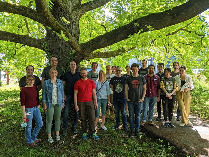 The Knop Lab team (NOTE: Photo taken with respect to social distancing measures and COVID regulations within Germany.)
“My focus is on method development,” Krisztina continued. “Currently, I am working on optimizing a PCR tagging method, a one-step procedure for chromosomal gene tagging that enables the rapid creation of mammalian cell lines with targeted large chromosomal insertions, such as green fluorescent protein (GFP). These proteins can then be visualized with a microscope or cell imaging system. The aim is to establish a universal, scalable and high-fidelity method to tag proteins in mammalian cells, enabling researchers to tag and target as many proteins as possible.”
The group’s PCR tagging method involves the transfection of a gene-specific PCR cassette into the target cells, together with a helper plasmid containing a Cas12a endonuclease, fusing a tag – such as GFP – onto the gene of interest. The process uses two gene-specific tags – termed M1 and M2 – with homology arms ranging from 50 to 90 nucleotides for targeted integration by homology-directed repair-based (HDR-based) genome editing. The M2 tagging oligo also provides a protospacer sequence for the Cas12a endonuclease. The generic template cassette contains a Cas12a-specific crRNA gene consisting of a promoter and crRNA direct repeat, as well as providing the fluorescent tag and additional features, such as a selection marker. Expression of the crRNA gene inside the cell directs Cas12a expressed from the helper plasmid to a target locus close to the insertion site, causing a double strand break. The linear tagging cassette is then incorporated into the genome at this site by HDR. The homology arm of the M1 tagging oligo directs in-frame fusion of the tag with the target open reading frame, leading to the expression of a tagged protein.
Krisztina continued: “When developing this universal method for CRISPR-Cas12a-assisted PCR tagging of mammalian genes, we initially relied on fluorescence microscopy methods for live cell imaging, and still use this technique extensively. However, we wanted a more scalable approach, and so acquired a Spark® Cyto, a multimode plate reader with fluorescence imaging and cytometry capabilities. This system is a great addition to the lab, giving us another way to gather information about the cells, and the success of the fluorescent tagging process. It allows us to rapidly image mammalian cells in a high quality and reliable way, and is ideal for optimization studies, as it can simultaneously image the entire microplate. This is a real advantage if we have several set-ups to evaluate, as we can image every well used in the study. It’s a much faster way to analyze cells than microscopy, because it not only enables live cell imaging, but also allows us to take advantage of the Image Analyzer™ software package to immediately quantify the success and efficiency of tagging experiments. Most of the time, we’re working with a single fluorophore at this stage, but we can also use the Spark Cyto’s multicolor application to automatically analyze multiple channels, to perform colocalization studies or introduce additional labels.”
It’s a much faster way to analyze cells than microscopy, because it not only enables live cell imaging, but also allows us to take advantage of the Image Analyzer software package to immediately quantify the success and efficiency of tagging experiments.
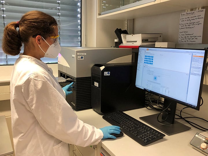 The Knop Lab is taking advantage of the Spark Cyto’s fluorescence imaging and cytometry capabilities capabilities for PCR tagging experiments
“Although we mainly use the Spark Cyto for end-point measurements, we also have the option to incubate cells inside the reader if necessary. Most experiments only take a couple of minutes per plate, but it’s good to know that we can incubate the cells in the measurement chamber if we need to, to make sure that they remain viable throughout the experiment. As well as controlling the reader temperature at 37 °C, we also plan to take advantage of the integrated gas control function to maintain levels of CO2 in the future. It’s been an interesting journey getting to know the Spark Cyto and discovering what we can use it for, and we’ve enjoyed excellent support from Tecan,” concluded Krisztina.
Keywords: