Stem cell research has seen explosive growth in recent years, with the technology holding promise for the treatment and cure of a wide range of conditions, from cancer, diabetes and heart disease to neurological conditions, inherited disorders and conditions of aging, including age-related macular degeneration (AMD). The Stem Cell Institute (SCI) at the University of Minnesota, founded in 1999, was the first integrated stem cell institute to be established in an academic environment, and focuses on basic and translational research with these versatile cells.
The ability of stem cells to differentiate into a variety of other cell types offers the potential to treat and cure a range of diseases and benefit patients worldwide. The University of Minnesota SCI is a collaborative center for researchers from 25 university departments, focused on gaining a greater understanding of stem cell biology and the potential uses of this technology. It has an emphasis on the study of induced pluripotent stem cells (iPSCs), primarily because they can be generated from any individual, and can be used to model diseases, discover patient-specific drug responses and potentially generate autologous cells for transplant therapies.
Dr James Dutton, Associate Professor at the SCI and the Department of Genetics, Cell Biology and Development, and Director of the SCI Innovation Facilities, explained: “My group researches an array of diseases that currently have no cure or effective treatment. One major focus is dry AMD, a disease affecting older individuals that causes patients to lose their central vision. This condition is highly prevalent in the Northern European population that settled in Minnesota, and I work closely with Professor Deborah Ferrington from the Department of Ophthalmology and Visual Neurosciences to investigate this area. Our studies center on mitochondrial dysfunction in the retinal pigment epithelium (RPE) – a single cell layer at the back of the eye – that is damaged in the eyes of AMD patients. Much of the initial work was conducted using eyes donated to the Lions Gift of Sight eye bank, however, to move this research to our AMD patient population, we needed to find a way to look at RPE cells from living individuals. We have done this by taking a 2 mm biopsy of the conjunctival layer covering the white part of the eye, reprogramming these cells to produce iPSCs that then undergo a differentiation protocol that causes them to become RPE cells. Using these iPSC-RPE cells, we are able to screen a range of potential candidate drugs, aiming to find effective ways of maintaining or improving mitochondrial function.”
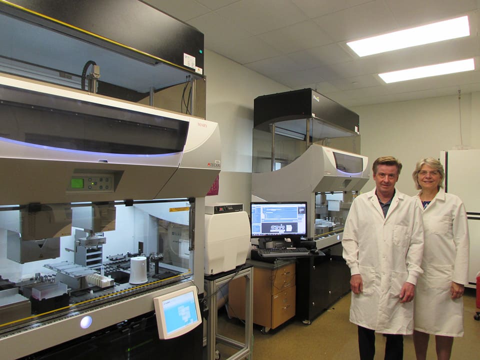
 Associate Professor James Dutton and Professor Deborah Ferrington in the dedicated stem cell laboratory
“Scaling up this process of making iPSC-derived RPE and testing the cells in a targeted drug screen would be extremely difficult without automation; we would need a large team of people, and it would be very slow and prone to error. However, until recently, automation of iPSC derivation, culture and differentiation has been complicated and technically challenging. We needed to find a user-friendly, cost-effective automation platform that would perform these methods efficiently, demonstrate scalability and commercial viability, and would fit in the space we had available.”
“With support from a generous philanthropic donor, we looked at a number of options and chose two Fluent® 780 platforms, and the department refurbished laboratory space specifically to house these units. The systems are excellent; we can perform the whole end-to-end process of derivation, culture and differentiation of the iPSCs into RPE and set up for the drug screening process on them. We designed the systems to suit our needs, including an integrated HEPA filter hood to create a sterile environment, a LiCONiC automated incubator to hold the plates, a Cytation™ 1 Cell Imaging Multi-Mode Reader to monitor cell confluence and morphology, and an integrated centrifuge for spinning down cells during passage. Additionally, there are two EchoTherm™ IC20 dry baths on the deck for sample and media temperature control, and a barcode reader to track samples throughout the process. The platform has three arms: an eight-channel Liquid Handling Arm™, a MultiChannel Arm™ 384 and a Robotic Gripper Arm™, which together, are able to perform all tasks associated with cell handling. The Tecan Labwerx™ Group has been especially good at ensuring full connectivity between all the equipment from third-party manufacturers.”
“The platforms are very intuitive to use and can replicate everything that we used to do by hand – we can even control the speed, pressure and pattern of pipetting. We can measure colony size and cell density to track growth using the integrated imager, and the systems automatically perform daily media changes at the differentiation stage when the iPSCs are converted into RPE cells, saving us hours of ‘people power’. The Tecan team is now refining its scheduling software to further complement our system, so that we can connect our entire protocol and minimize the need for human input. This will increase efficiency and, using multiple platforms, the workflow could easily be scaled up to a commercial level if necessary.”
The platforms are very intuitive to use and can replicate everything that we used to do by hand - we can even control the speed, pressure and pattern of pipetting.
“We now have data from both donor eyes and living patients, and we are building up a patient cell bank of conjunctival cells, iPSCs and iPSC-derived RPE, as well as generating results from the preliminary drug screens. The aim is to add to our data over the next few years so that we can start to see what drugs can help AMD patients. At the same time, we are planning to automate a number of other differentiation protocols on the platforms to make other cell types from iPSCs, such as glial cells and neurons. From there, we would like to expand into more technically difficult procedures such as organoid and 3D cultures – it’s an exciting time,” concluded James.
To find out more about Tecan’s cell biology solutions, visit www.tecan.com/cellbiology
To learn more about the Minnesota Stem Cell Institute, go to www.stemcell.umn.edu
Keywords:


