


Keywords:
By Siegfried Sasshofer
Imagine life science research without cell-based assays. Or without cultured cells of all types to power those assays. Healthy, high-quality cells at the right point of confluence are vital for proliferation, kinetics, cytotoxicity, and gene expression studies particularly during long-term experiments. With so many different cell types, assay formats, and detection methods the variability inherent in cell-based assays can be enormous. There’s no room for inconsistency in cell counts and confluence assessments — it’s counterproductive and just wastes time. What’s the best way to improve counting accuracy in your cell-based assays?
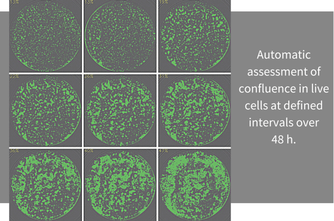
Automatic assessment of confluence in live cells at defined intervals over 48 h. Cells were maintained, imaged, and measured within Spark® automatic imaging cell counter.
Cell type governs all else
That’s what cell-based assays are all about. Error here can propagate through an entire experiment. Of course cell density analyses and quality checks are routine for cell seeding, passaging, and to assess the cell health before beginning cell-based assays.
Accurate cell counts are especially vital during live-cell kinetic assays and when measuring differences among samples is the objective of your studies. Your first step in ensuring consistency is simply to look at your cells — you recognize them and you know a healthy culture when you see it. Then count the cells using the most accurate, precise, and reliable method available to you.
Environmental control governs optimal cell confluence
Take care of your cells. Give them what they need, then monitor viability in culture and you’ll know they’re in good shape for your cell-based assays. Make sure your temperature, humidity and gas concentrations are as accurate and as stringent as your most sensitive cells require. But when you run cell-based assays, you face a dilemma. Just when you need your cells to be on their best behavior, they must come out of their safe environment to get ready to run.
Your cells are at greatest risk during experiments. For cells in vivo, environmental and mechanical stress can be part of everyday life. But cells in culture are far more delicate. Even small changes in temperature, gases, and mechanical shock may induce behavioral responses, without necessarily causing any obvious physiological damage. For more consistent cell counts, protect and monitor your cells for as long as possible both in culture and during assays.
Experimental variability governs data quality
Each step you take to improve precision in cell counting and consistency in confluence assessment, to protect cell health, and to minimize edge effects reduces experimental variability in live-cell kinetic assays. Statistical analysis is simplified. You can limit replicates to only those you need to confirm your experimental conclusions, rather than running extras to average out experimental error. You spend less time counting cells, your results are clearer and you’re left with only the performance differences that will enable you to draw the insights that will conclude your studies.
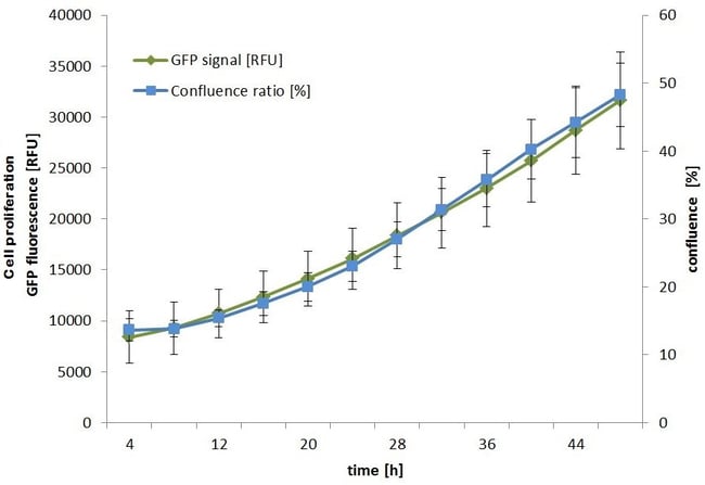
The live-cell kinetics assay conducted in the same culture as above reveals nearly identical kinetics in cell growth (GFP signal) and confluence ratio at intervals over 48 h. Fluorescent signal and confluence were measured simultaneously on Spark® automatic imaging cell counter.
Analyzing true differences among experimental conditions is the core of cell-based assays. That’s where the excitement is. Why complicate your experiments with added variability from cell counting, confluence assessment, and assay stress? Diagnosing and minimizing each source of error can be tedious and time-consuming. With a multi-functional automated imaging cell counting system you can leave the tedium of experimental error behind, and use your time for the more exciting results you seek.
Want to know more?
Download this application note that describes a method for walk-away monitoring of cytotoxicity, viability and apoptosis assessment.

About the author

Siegfried Sasshofer
Siegfried Sasshofer heads the marketing team responsible for Tecan’s Detection products based out of Tecan’s development and manufacturing center for microplate readers and washers in Grödig, Austria. He has over 20 years’ experience in working in the field of bio-analytics. He studied Biotechnology, holds an MBA and joined Tecan in 1997.