


By Dr Stefan Haberstock
Cell-based assays are giving us deeper insight into cellular mechanisms in a true biological context, and fluorescence assays are playing a leading role. Applications range from cytotoxicity, proliferation, apoptosis and G-protein-coupled receptor (GPCR) signaling assays to high-throughput screening (HTS) drug discovery.
When you are developing a cell-based fluorescence assay you will need to handle issues common to cell-based assays in general, such as tight environmental control, avoidance of microplate edge effects, and cell density optimization. There are also issues specific to fluorescence assays to address, including selection of dyes and optimization of excitation and emission wavelengths that we looked at in the previous article in this series. Finally, the cell culture environment itself also raises challenges that can be overcome with the right approach, as we will see here.
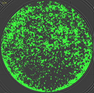
Cell-based fluorescence assays have revolutionized our view of cellular processes. This image shows automatic cell confluence determination using Spark's integrated brightfield optics and software based labeling (green) of identified cells.
How to cope with autofluorescence background
Autofluorescence is a typical problem that needs to be addressed when developing a high-performance cell-based fluorescence assay. A common source for this background fluorescence is the phenol red used as pH indicator in most cell media. One strategy to minimize the influence of the phenol red on cell-based assays with adherent cells is to read from the bottom. This avoids that light has to travel through the medium on the way from fluorophore to detector.
Choosing the wrong plate can also lead to higher background. White plates increase the background in a non-time resolved arrangement because they reflect the light used for excitation. Clear plates, on the other hand allow cross talk between neighboring wells. Black plates with a transparent bottom are therefore always the first choice in cell-based assays with adherent cells, since they minimize background, inhibit crosstalk and allow bottom reading.
Advanced optics enable analysis of cell monolayers
Detecting and measuring fluorescence in a homogeneous liquid sample is one thing, but cell-based assays generally involve analyzing adherent cells that are unevenly distributed across the bottom of the well. Achieving a robust sensitive assay therefore means covering the well bottom with multiple measurement points to analyze the whole well bottom. Added to that, achieving high assay sensitivity depends on measuring signal only from the thin monolayer of adherent cells. Focusing the emission detection to the well bottom maximizes signal and minimize background fluorescence, for example from the column of medium above the monolayer. Together, these requirements demand sophisticated optics that enable emission detection to cover the complete well bottom and focus signal detection on a narrow depth of the well volume.
Signal intensities from cell assays greatly depend on the cell number in each well. To ensure that results can be compared, the cell number should be checked before starting the experiment and also before detecting the signal. This enables the signal to be normalized and easy identification of outliers caused by wells with deviating cell numbers.
Capabilities for demanding cell-based fluorescence assays
Implementing fluorescence detection is certainly one of the easiest and safest ways for you to improve the quality and sensitivity of your assays. In the final article in this series, we will look out how the Tecan Spark multimode microplate reader can deliver the flexibility you need to develop a wide range of fluorescence-based biochemical and cell-based assays, for your needs now and in the future.
For more information on measuring cell confluence to improve measurement consistency, get the application note: Accurate and Automated Cell Confluence Assessment in Microplates.

About the author

Dr Stefan Haberstock
Dr Stefan Haberstock works as a sales development specialist for benchtop solutions at Tecan with over 5 years in experience in field marketing and supporting product launches. He studied Biology at Gutenberg University Mainz and Trinity College Dublin and received a PhD in Biochemistry from Goethe University Frankfurt am Main.