


Discovering and developing new antimicrobial drugs to tackle antibiotic resistance requires an understanding of how bacteria respond and adapt to new compounds. A range of tests are needed to determine the efficacy of potential drugs, such as aggregation and adhesion/invasion assays. For SMALTIS, a biotechnology company in Besançon, France, test automation has dramatically improved throughput and data collection, freeing up research hours to concentrate on developing new experiments.
SMALTIS, established in 2014 by Dr Sophie Guénard and Dr Cédric Muller, is a CRO specializing in microbiology, molecular and cellular biology. It off ers services to private and public research laboratories, assisting with R&D projects by providing customized bacterial strains, evaluating the activity of antimicrobial compounds and analyzing microbial samples. The company was born out of a shared research background, as Cédric explained: “Sophie and I studied microbiology at the Patrick Plésiat labs, seeking to understand the antibiotic resistance mechanism exhibited by Pseudomonas aeruginosa, the primary cause of death in cystic fi brosis. We were fortunate to work with a big pharma company to develop a specifi c antibody vaccine and, thanks to the project, we got the idea of creating a company dedicated to helping laboratories accelerate their microbiology R&D. We perform minimum inhibitory concentration (MIC) studies and time-kill experiments, characterizing how the test compounds act on the target bacteria, and how the bacteria in turn adapt to the drug, helping our clients to improve the effi cacy of their compounds.”
Aggregation and adhesion/invasion assays are among the many services SMALTIS off ers. In order to colonize and initiate an infection, bacteria adhere to host cells via the fi mbriae (hair-like structures) that interact with specifi c host cell receptors. Aggregation assays are used to assess the ability of a compound to inhibit adhesion, by interacting with the fi mbriae to cause aggregation. Cédric said: “Our basic assay monitors drug-bacteria interactions by taking regular images over the course of fi ve hours. When we started performing aggregation assays, our customer gave us a specifi c operating procedure which was designed for a high content imaging platform. As these systems are expensive, we looked at alternative approaches, and developed a manual set-up: we incubated the plate on a rotating incubator and removed it every hour for analysis, then returned the plate to the incubator. Although this worked well, it was very labor intensive.”
“We then came across a Spark® multimode microplate reader in one of the research laboratories of Jean-Minjoz Hospital that we collaborate with. We developed an aggregation assay protocol on the hospital’s instrument, and a member of the Tecan team helped us to perform some validation experiments to confi rm that we could transfer our technology and methods onto the system. We then went ahead and purchased our own Spark reader, allowing us to perform the same aggregation assay far more costeff ectively than using a high content imaging platform.”
Cédric continued: “The experiment now runs without the need for supervision, with the added benefi t that the Spark produces growth curves of the strains over the course of the assay. Automation has freed up our time to develop other experiments, for example, we are currently developing a method to evaluate luminescent bacterial strains, created by inserting a copy of the luxCDABE operon – from Photorhabdus luminescens – into their DNA.”
"What took one person three days, now takes an hour and a half."
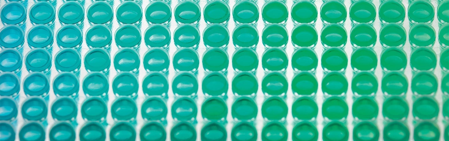
“We also use the Spark to carry out adhesion/invasion assays to determine the capacity of bacterial strains to attach to or invade eukaryotic cells. The cells are infected with a bacterial strain, then after a series of washes, the eukaryotic cells are stained with DAPI and the bacteria with specifi c antibodies conjugated to an AlexaFluor® dye. We measure the blue and green fl uorescence, from the eukaryotic cells and bacteria respectively, to determine a ratio refl ecting the strain’s adhesion/invasion capabilities. Prior to having the Spark, we had to take four to six images to cover each 96-well plate, and analyze the colours with a microscope. This is now done automatically, and what took one person three days, now takes an hour and a half. We are very happy with the Spark; it easily adapts to each assay set-up, and its high throughput and automation has enabled us to continually improve our service for our clients,” Cédric concluded.
All Tecan products mentioned are for research use only. Not for use in clinical diagnostics.
Keywords: