


By Dr Stefan Haberstock
Compared to many other detection technologies, fluorescence provides hard-to-beat performance and flexibility. Fluorescent labels are stable for months, deliver high sensitivity and the diversity in available dyes gives nearly unlimited possibilities in assay design. This and many other advantages make implementing fluorescence detection one of the easiest and safest ways for you to improve the quality and sensitivity of your assays.
Stable strong signal
Fluorescence has established itself as the major detection technology in the biosciences for many reasons. Fluorescence does not require addition of detection reagents and can be read multiple times, essentially as often as the sample can withstand exposure to light. Some fluorescent dyes, like the Lanthanides, have special properties including long decay times of the emission signal. Measuring the emission of lanthanides in a time resolved manner with a delay of a few hundred microseconds increases signal to noise and boosts sensitivity.
Fluorescently labeled reagents are also highly stable, with long shelf life that enables efficient preparation of large standardized batches that can be used for extended periods. This minimizes inter-assay reagent variability.
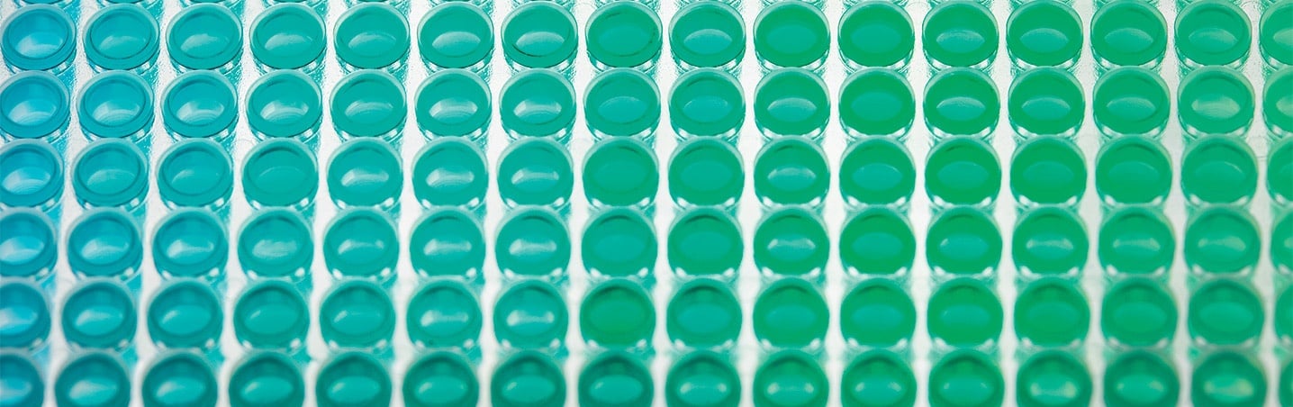
Fluorescence-based assays are stable, simple and sensitive
Seemingly endless possibilities
The enormous popularity of fluorescence-based detection has stimulated the development of a myriad of kits from a large number of suppliers that ensures fast access to research tools. Fluorescent tags can be added to almost every biomolecule and can be used for many applications, such as identification, quantification or tracing biological interactions. Some suppliers even offer a choice of biomolecules already labeled with a fluorophore, ready to use. Other commercially available fluorescent molecules include nucleotides and enzyme substrates.
The development of molecular biology based fluorescence labeling has revolutionized the study of gene expression. For example, Green Fluorescent Protein (GFP) can be fused to proteins or used as a reporter to follow gene expression, enabling researchers to study processes in living cells that were previously invisible. GFP fusion is also increasingly popular in protein biochemistry to monitor protein production and purification without the need for chemical labeling.
Multiplex for normalization and multi-target identification
Using fluorescence detection, you can measure a number of target molecules in a sample simultaneously by labeling each with a different fluorophore, and this ability to multiplex can make fluorescence the technology of choice in many situations. For example, you can use fluorescence to measure the numbers of live cells and dead cells in a sample simultaneously, by staining one red and the other green. Multiplexing is powerful because it allows researchers to interrogate cells of a given state on multiple targets. All you need is a system that can flexibly accommodate a variety of detection settings and is able to deliver high sensitivity over the complete spectral range.
From optimizing a fluorescence assay to final analysis
In the next two articles in this series, we will look at meeting the challenges in establishing a fluorescence assay, specific issues in the fluorescence-based study of cells, and how to choose instrumentation for fluorescence analysis.
Sign up to the blog to receive updates!

About the author

Dr Stefan Haberstock
Dr Stefan Haberstock works as a sales development specialist for benchtop solutions at Tecan with over 5 years in experience in field marketing and supporting product launches. He studied Biology at Gutenberg University Mainz and Trinity College Dublin and received a PhD in Biochemistry from Goethe University Frankfurt am Main.